By the middle of the 19th century, surgeons were trained with the use of atlases, which depicted the systems of human organs separately from each other. Anatomists drew organs after cadaver dissection — for this purpose they had to open cavities, destroy connective tissue, as well as remove fiber. This approach caused the change of mutual disposition of organs.
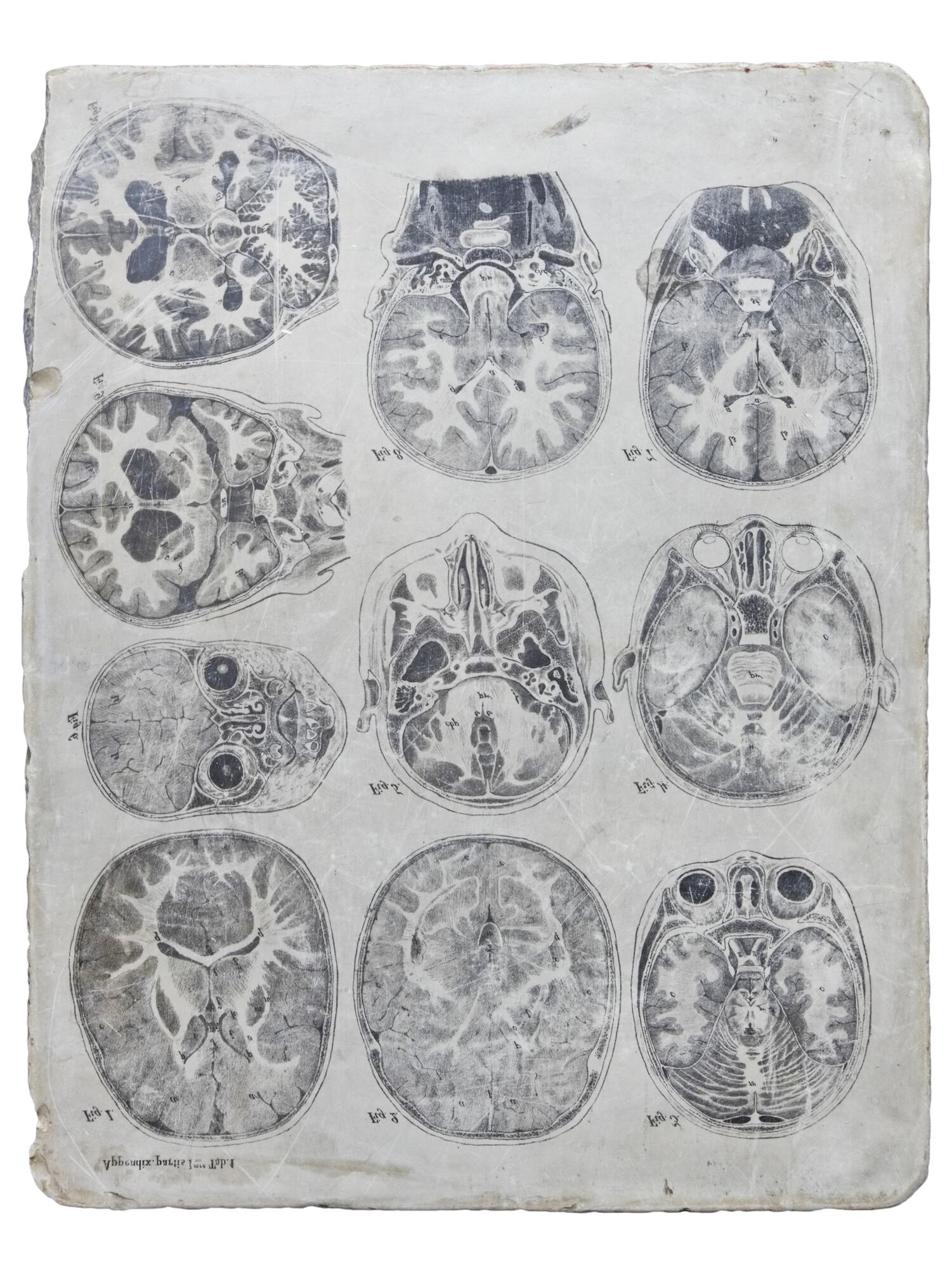
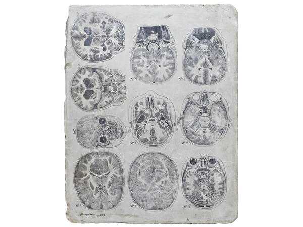
Based on his practical experience, the Russian surgeon and scientist Nikolay Pirogov came to the conclusion that it is not suitable to train specialists using such atlases. While working at the Anatomical Institute under the St. Petersburg Medical-Surgical Academy, he suggested a new method to study the exact location of organs and tissues in the human body. Pirogov froze fresh cadavers at a temperature of -25°C to the density of wood. It took 2-3 days. Then, using a special saw brought from a carpentry factory, the scientist sawed the frozen corpses into parallel plates several millimeters thick.
The cadaver was cut in three projections: along the body, dividing it into anterior and posterior sections (frontal projection), across the body (horizontal) and along the body, dividing it into left and right halves (sagittal). The images from the cut plates were covered with scale pattern glass and then redrawn in full size on scale paper. By combining and comparing the images, a surgeon could get a full picture of the organs and systems disposition to each other. During the operation, a doctor mentally imagined frontal, horizontal and sagittal cuts made through one point or another.
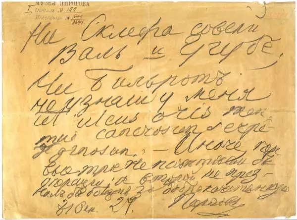
Pirogov wrote about these saw cuts in his autobiography:
Based on his practical experience, the Russian surgeon and scientist Nikolay Pirogov came to the conclusion that it is not suitable to train specialists using such atlases. While working at the Anatomical Institute under the St. Petersburg Medical-Surgical Academy, he suggested a new method to study the exact location of organs and tissues in the human body. Pirogov froze fresh cadavers at a temperature of -25°C to the density of wood. It took 2-3 days. Then, using a special saw brought from a carpentry factory, the scientist sawed the frozen corpses into parallel plates several millimeters thick.
The cadaver was cut in three projections: along the body, dividing it into anterior and posterior sections (frontal projection), across the body (horizontal) and along the body, dividing it into left and right halves (sagittal). The images from the cut plates were covered with scale pattern glass and then redrawn in full size on scale paper. By combining and comparing the images, a surgeon could get a full picture of the organs and systems disposition to each other. During the operation, a doctor mentally imagined frontal, horizontal and sagittal cuts made through one point or another.
Pirogov wrote about these saw cuts in his autobiography: